
NCI-H460-Luc
CBP30033L
| I. Introduction | |||||||||||
| Host Cell: | NCI-H460 | ||||||||||
| Expressed gene: | Luciferase | ||||||||||
| Stability: | 32 passages(in-house test, that not means the cell line will be instable beyond the passages we tested.) | ||||||||||
| Freeze Medium: | 90% FBS+10% DMSO | ||||||||||
| Culture Medium: | RPMI-1640+10%FBS+2μg/ml Puromycin | ||||||||||
| Mycoplasma Status: | Negative | ||||||||||
| Storage: | Liquid nitrogen | ||||||||||
| II. Description of Host Cell Line | |||||||||||
| Organism: | Homo sapiens,human | ||||||||||
| Tissue: | Lung:pleural effusion | ||||||||||
| Disease: | Carcinoma;large cell lung cancer | ||||||||||
| Morphology: | Epitheloid cell | ||||||||||
| Growth Properties: | Adherent | ||||||||||
| III. Representative Data | |||||||||||
 |
|||||||||||
|
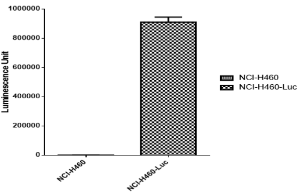
Figure 1. Detect Luciferase assay by Promega Bright-Glo Luciferase Assay System.. |
|||||||||||
| IV. Stablity Test | |||||||||||
 |
|||||||||||
|
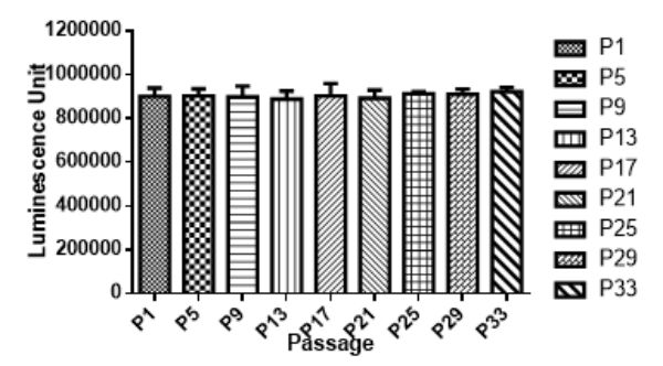
Figure 2. Stability test results. NCI-H460-Luc cells showed at least 32 passage stability with similar luminescence unit. |
|||||||||||
| V. In VIVO Data | |||||||||||
Intracranial injection:
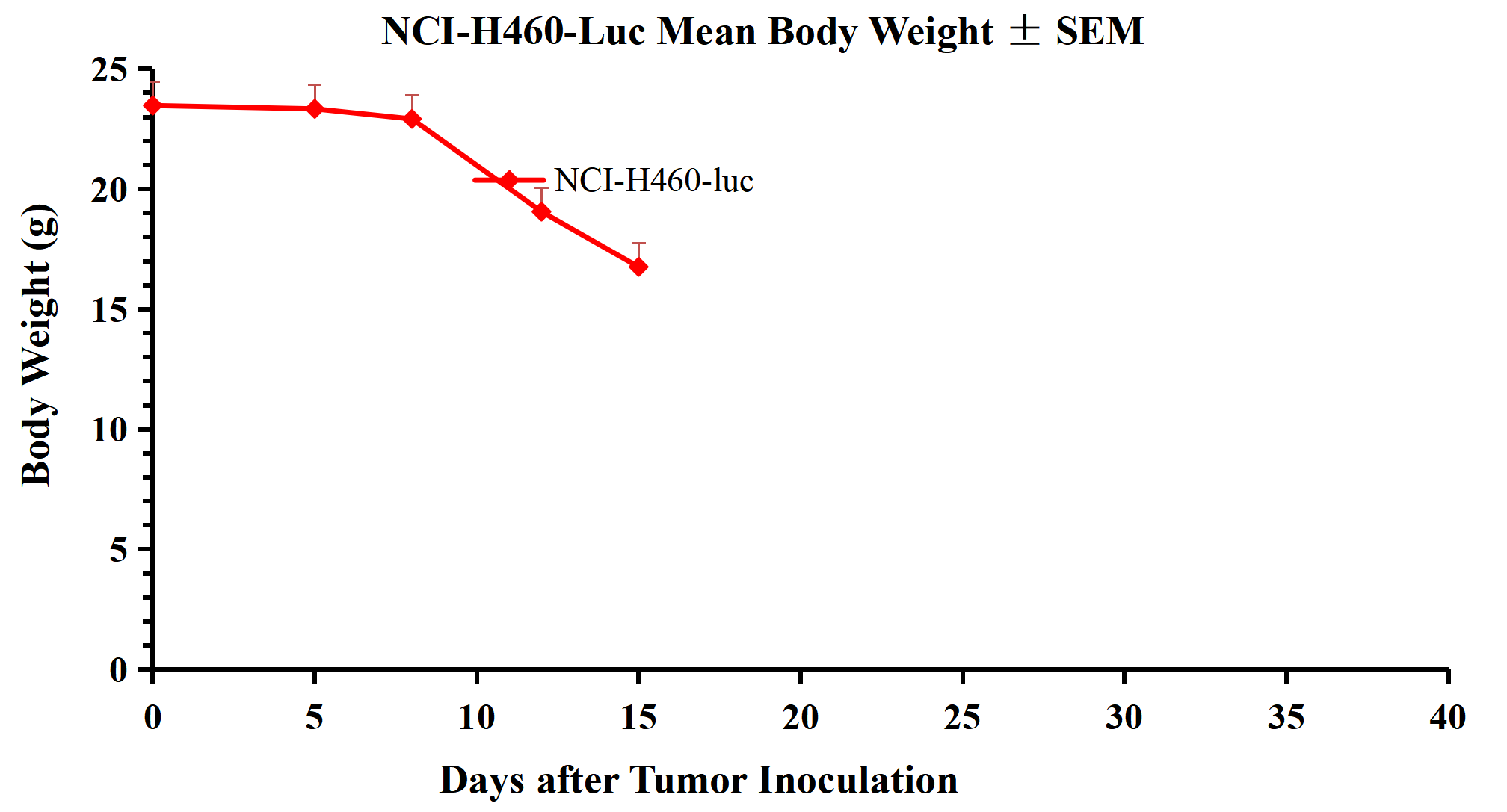
1. Weight Change 2. Total Flux
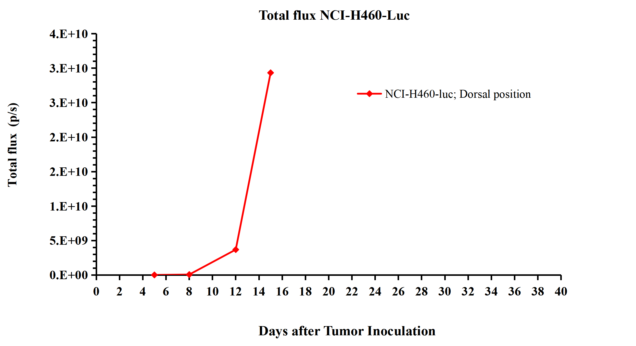 Figure 4. Total flux NCI-H460-Luc. 肺原位: |
|||||||||||
|
Index |
Content |
|
Animal |
BALB/c-nude |
|
Implanted Position |
肺原位 |
|
Injected Cell Numbers |
5×10^6/50μL |
|
Matrix gel |
Without matrix gel |
1.Tumor weight
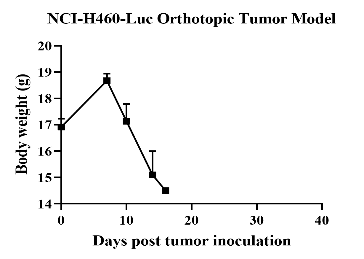
Figure 5. NCI-H460-Luc Orthotopic Tumor Model.
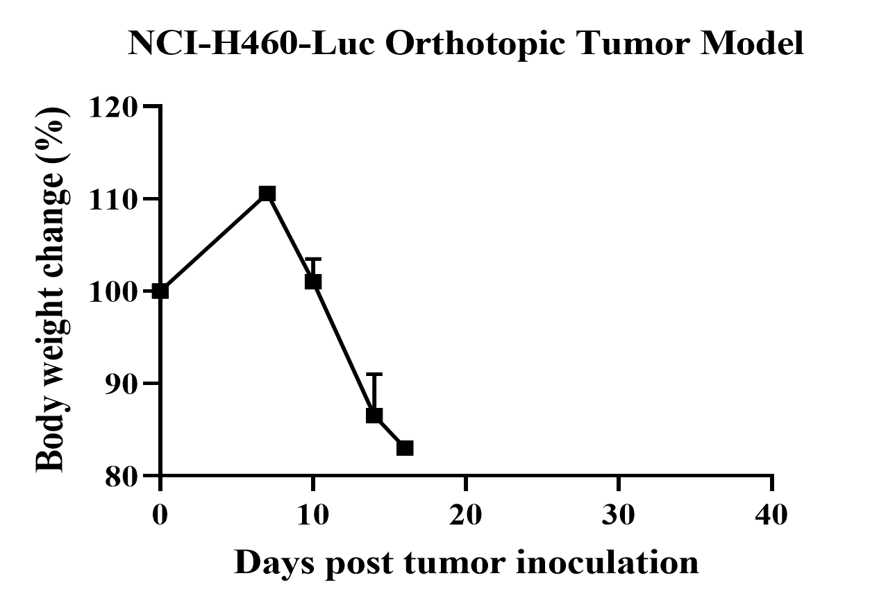
Figure 6. NCI-H460-Luc Orthotopic Tumor Model.
2. Tumor flux
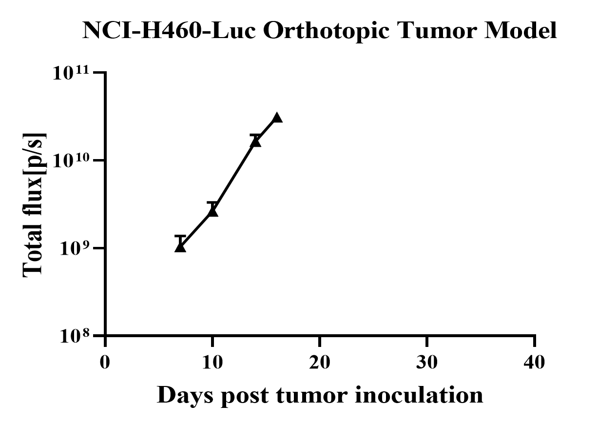
Figure 7. NCI-H460-Luc Orthotopic Tumor Model.
NOTE:The above data was provided by Cobioer (cell data) and Truway Bio (in vivo data model).